IRANDERMA |
|
Quiz: March 2006 |
This one-year-old baby was referred for a solitary nodular lesion that he has had since 4 months of age. He had not any other significant medical problem. Skin biopsy showed an infiltration of closely packed cuboidal cells in the dermis.
What is your diagnosis?
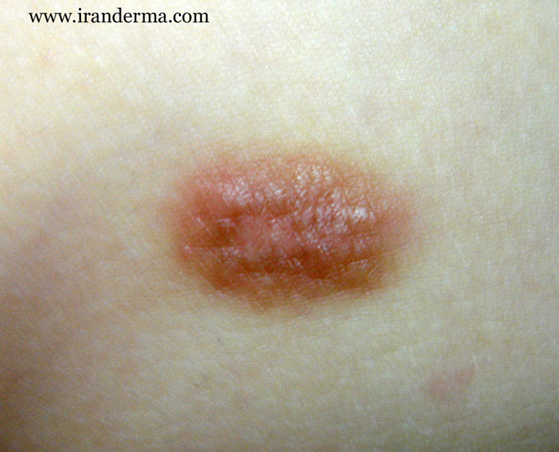
Diagnosis: Solitary mastocytoma
Comment by; M. Mehravaran, MD, Dermatologist/ Szeged, Hungary:
The
term mastocytosis encompasses a group of clinical disorders that
result from an abnormal proliferation of tissue mast cells in
variety of tissues in particular in skin. Mastocytosis occurs in
all races and affects both sexes equally. The peak incidence of
mastocytosis is in children, in over half of all patients the
disease is diagnosed before the age of 6 months (as in
Quiz case). There is second peak of incidence in young adult.
Mastocytosis tends to be transient in children and chronic in
adults
Two
subgorups of mastocytosis
patients are recognized: those with only skin involvement (cutaneous
mastocytoma) and those with mast cell infiltrates involving
several different organs (systemic mastocytosis).
Patients
with cutaneous mastocytosis can be generally classified
into:
- solitary
mastocytoma,
- urticaria
pigmentosa,
- diffuse
or erythrodermic mastocytosis,
- telangiectasia
macularis eruptiva perstans.
The
number of mast cells infiltrating the dermis in cutaneous
mastocytosis varies from a relatively small number that is
undetectable on the physical examination to larger aggregations
forming papules, nodules, or diffuse thickening of the skin. Papular
or nodular lesions with dense dermal infiltrates may
have a yellowish hue that accentuated by diascopy. Darier’s
sign, which is development of urtication and delayed,
axonally mediated erythematous flare, can usually be elicited by
rubbing or other minor trauma to a lesion. Such physical
stimulus causes mast cell degranulation with the release of mast
cell mediators and local tissue effects of vasodilatation,
increased vascular permeability, and edema. Generally other
changes include hyperpigmentation, flushing, localied or
generalized pruritus, occationally tense bullae in
infants. Unususal severe complications in infancy include
hypotension and shock, severe diarrhea and dehydration, and very
rarely a bleeding diathesis.
Mastocytoma
The
term mastocytoma has used to described nodular infiltrates (as
in Quiz case) of mast cells occurring single or one of several
isolated lesions. Solitary mastocytomas occur
almost exclusively in the first 2 years of life and are often
present at birth. These solitary nodules typically are found on
the trunk and extremities and range in size from 5-60 mm in
diameter. Darier’s sign, epidermal pigmentation, the formation
of vesicles and bullae, and localized flushing are common;
generalized flushing has rarely been reported.
Lesions
that are truly solitary usually regress completely or become
asymptomatic and rarely persist into adulthood.
Histologically
the infiltrating mass cells are usually monotonus and have round
or oval, darkly staining, bland nuclei and a moderate amount of
finely granular cytoplasm that gives the cells a distinctive
appearance like a fried egg.
Management
Most
solitary mast cell lesions resolve spontaneously, generally no
treatment is required. Therefore are self-limited and not-threatening,
reassurance of the usual benign course of this
disease and avoidance of specific factors known to trigger
mast cell degranulation such as immunologic (allergens),
physical (heat, cold, sunlight, trauma), biologic toxins (snake,
insect, jellyfish and shellfish) and drugs (aspirin, alcohol,
narcotics). Excision of symptomatic solitary
mastocytomas may rarely be indicated, e.g., if severe
cardiovascular or respiratory symptoms are being produced.
Since
no effective therapy exists for controlling mass cell
proliferation, treatment for the remaining subgroups of
mastocytosis patients is directed at inhibiting the local (Potent
topical corticosteroids) and systemic effects of released
mast cell mediators.
ايران درما |